Consultations
← Back to Consultations
← Back to Consultations
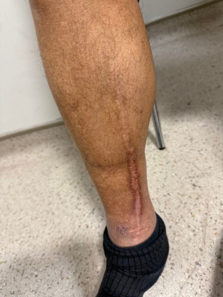
Pyoderma Gangrenosum
Pyoderma gangrenosum is a rare, painful, neutrophilic dermatosis causing rapidly enlarging necrotic ulcers, most commonly on the legs, and is a diagnosis of exclusion frequently associated with systemic disease.
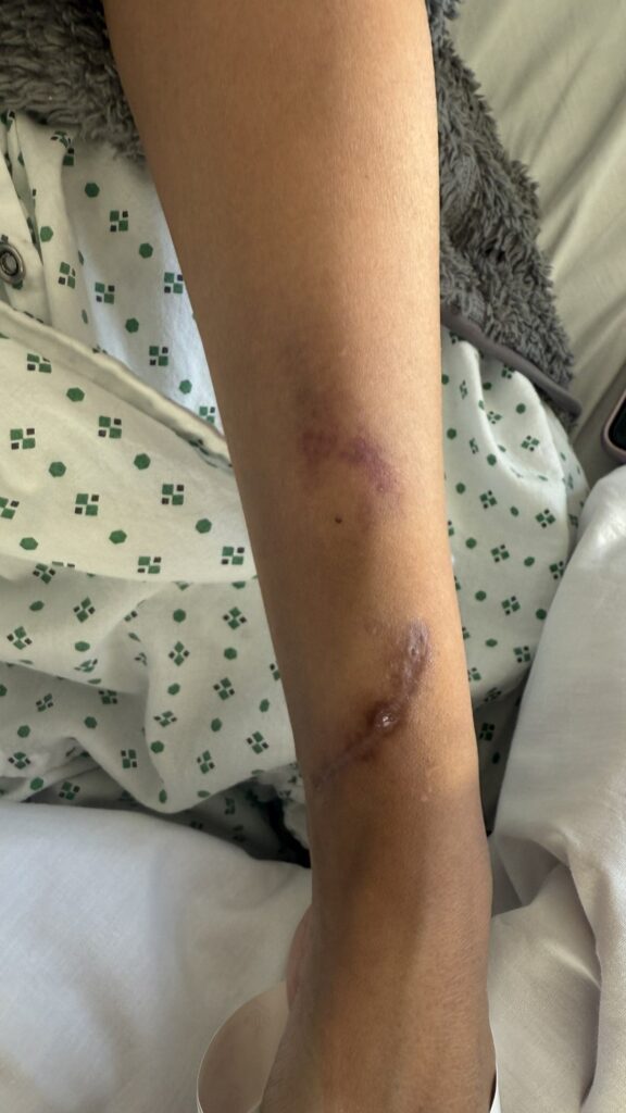
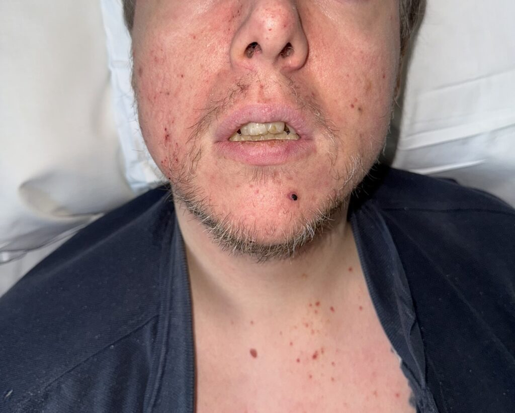
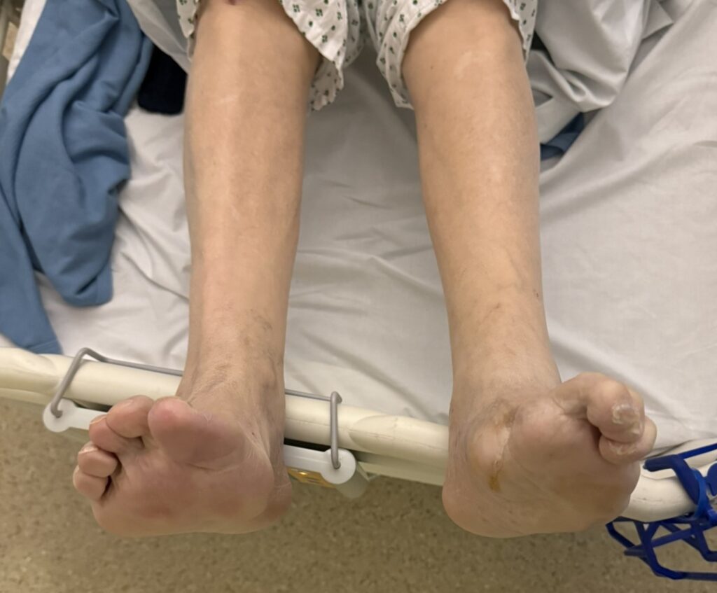
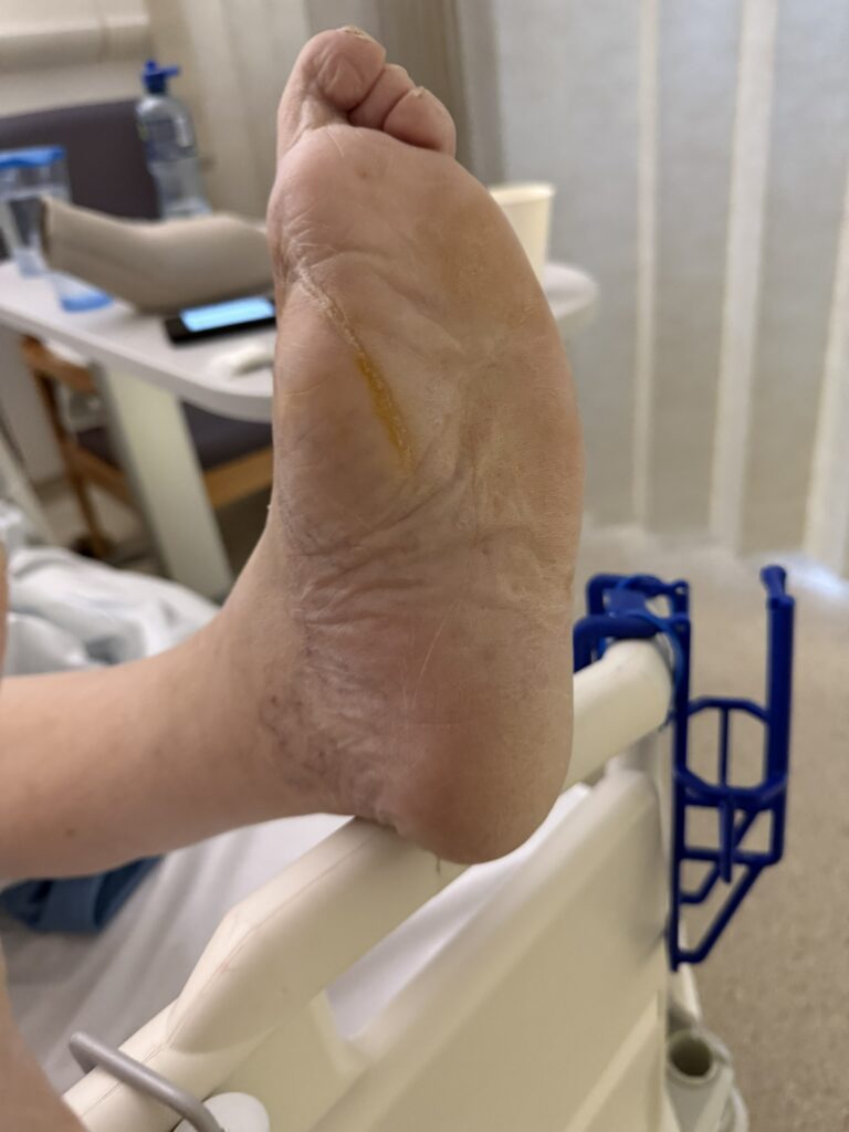
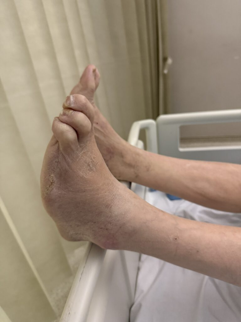
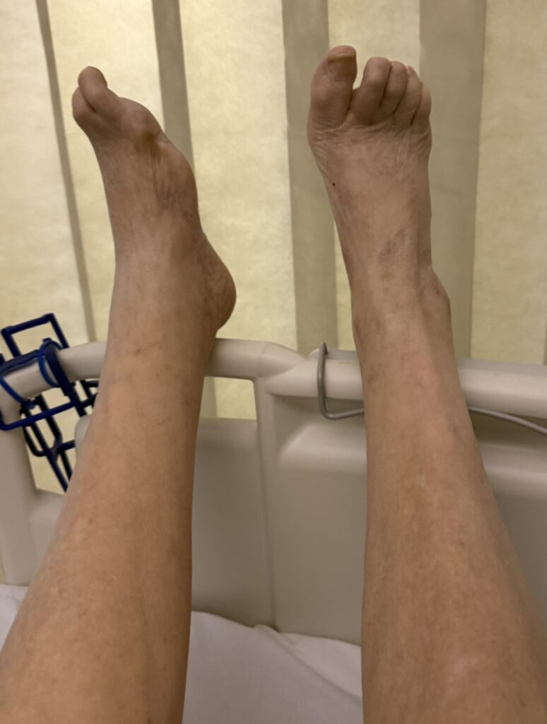
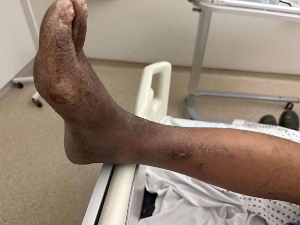
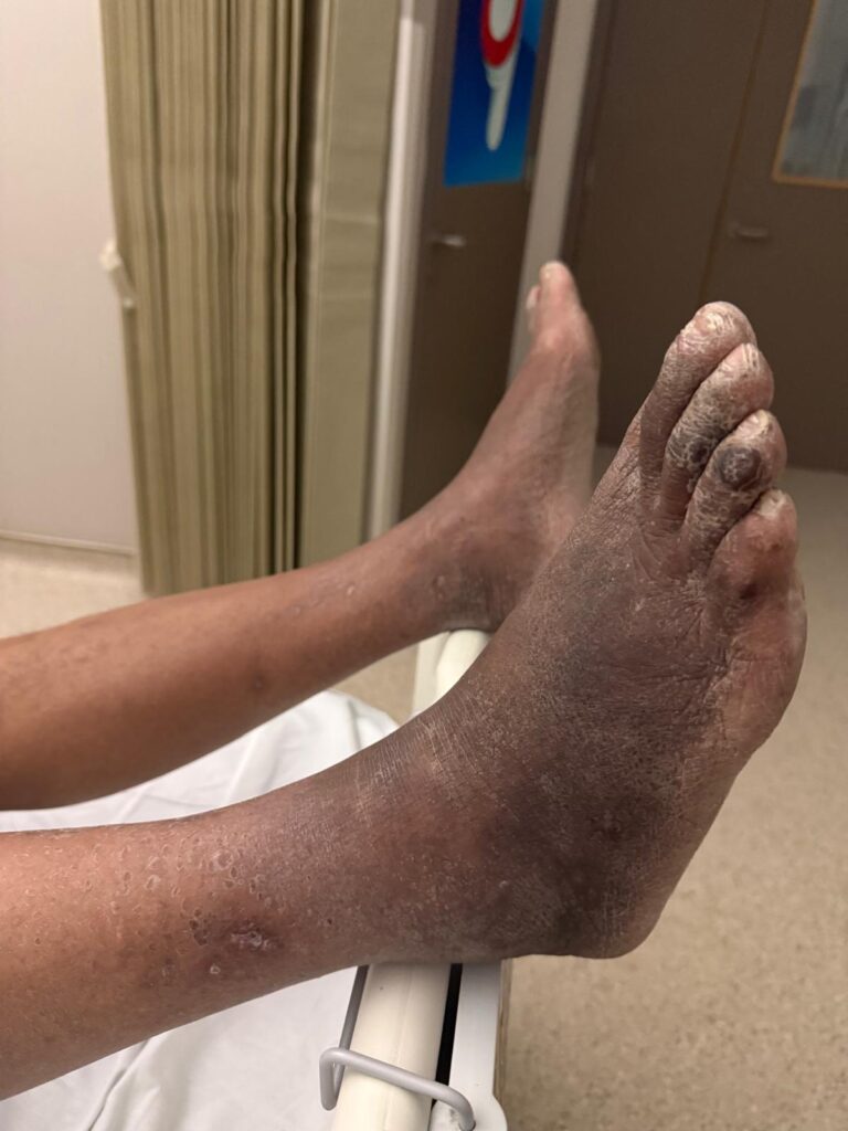
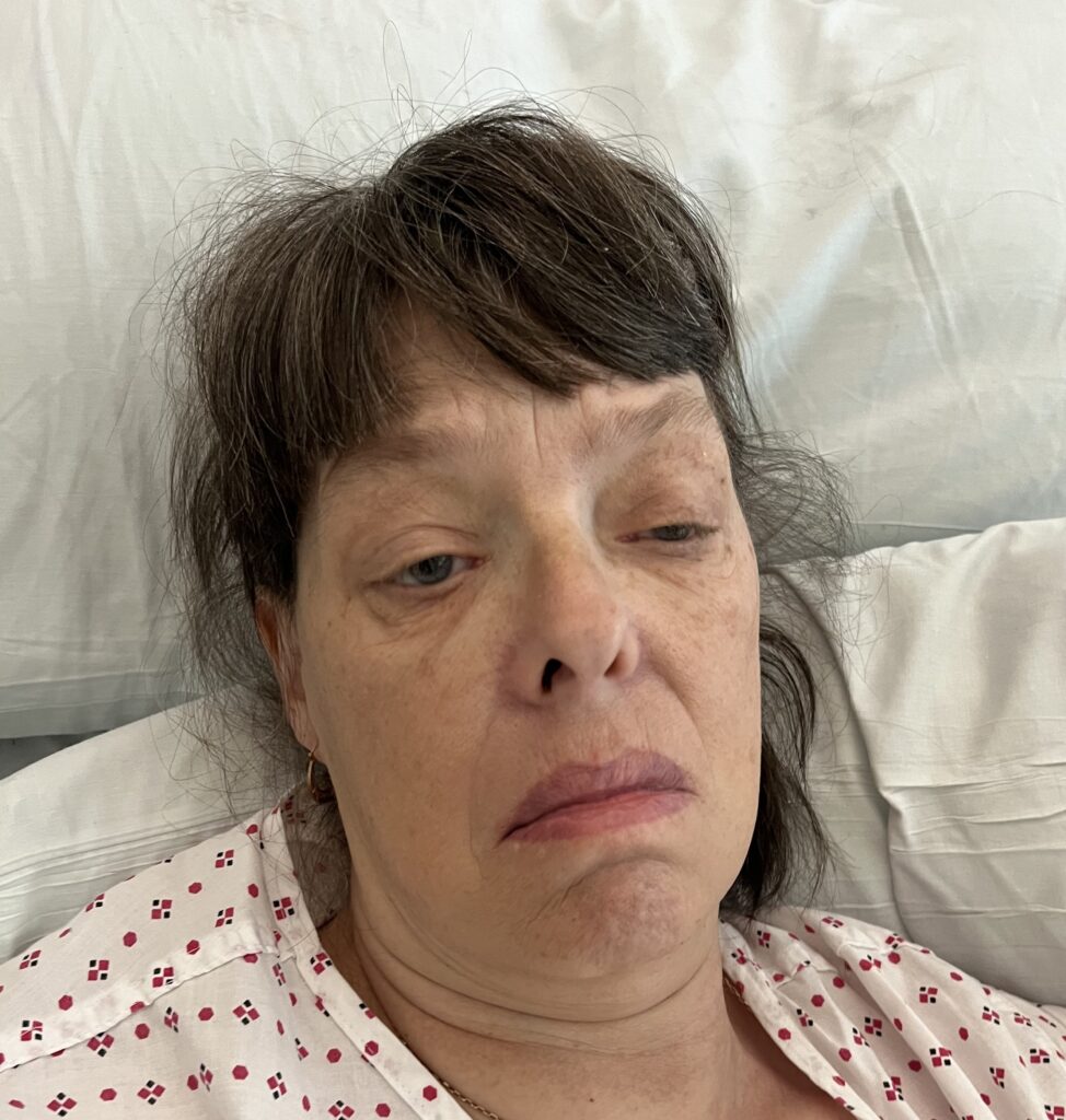
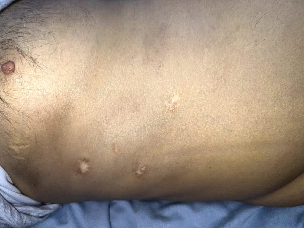
Station Instructions: Please assess this 55-year-old man with a non-healing leg ulcer on his left shin.
1. Key History-Taking Points
Timing & Progression
- Onset & duration: since when, sudden vs gradual onset, coming and going vs constant
- Progression: is the ulcer getting larger or more painful over time?
- Previous ulcers: any prior episodes here or elsewhere on the body?
- Initiating event: did it start after minor trauma? (pathergy is characteristic of PG)
Ulcer Characteristics
- Location & size: exact site, approximate dimensions
- Pain: severity, character — PG ulcers are typically very painful
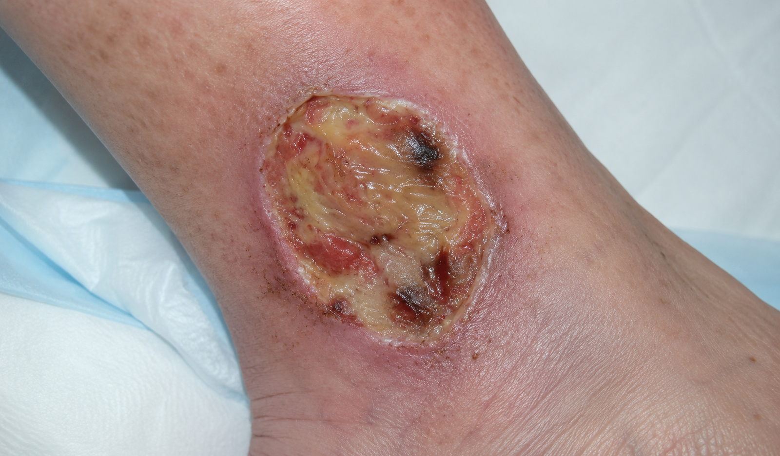
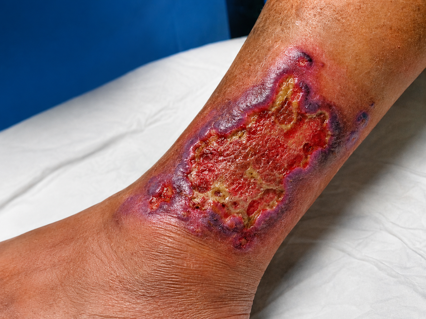
- Appearance: colour of the base, presence of black/necrotic tissue, purulent discharge, smell
- Edge: undermined, bluish-red, ragged margins are characteristic
Systemic & Associated Disease Screen
- Vascular risk factors: diabetes, hypertension, hypercholesterolaemia, smoking, prior MI/angina/stroke (arterial or diabetic ulcer)
- Neurological: pins and needles, numbness (neuropathic ulcer)
- Venous: varicose veins, DVT history, multiple pregnancies, prolonged standing
- Vasculitic & connective tissue: joint pains, skin rashes, mouth ulcers, dry eyes, Raynaud’s phenomenon, eye inflammation
- Gastrointestinal (IBD): bowel habit change, weight loss, abdominal pain, PR bleeding — IBD (especially UC) is the most common association
- Haematological malignancy: fever, drenching night sweats, lymphadenopathy, bleeding or bruising, back pain (lymphoma, leukaemia, myeloma)
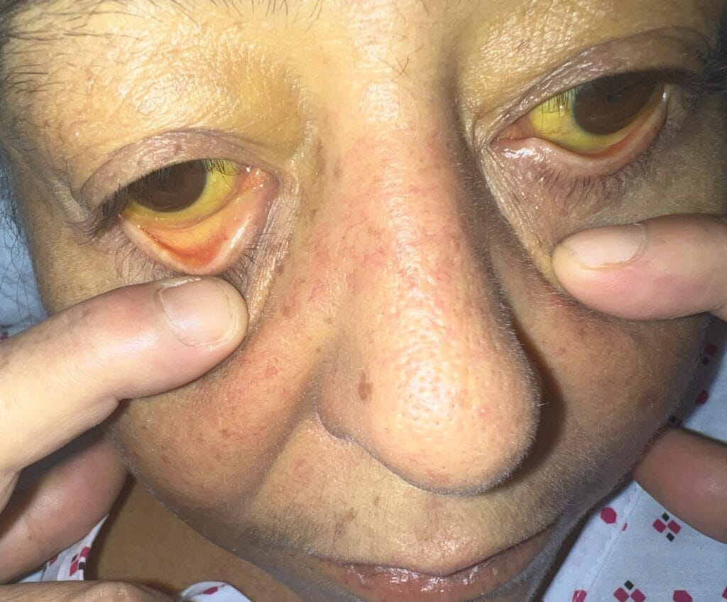
- Hepatobiliary: jaundice, itch (PBC/hepatitis C associated with PG)
- Fever: may indicate secondary infection rather than primary diagnosis
Past Medical, Drug & Family History
- PMH: known IBD, RA, haematological malignancy, diabetes
- Treatments tried: wound dressings, antibiotics, steroids — response to treatment is diagnostically informative
- Family history: IBD, autoimmune disease
- Social history: occupation, mobility, living situation
2. Key Examination Findings
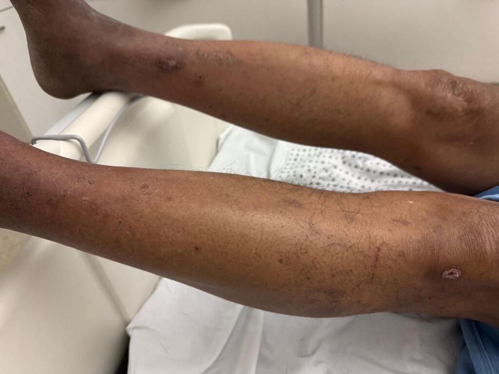
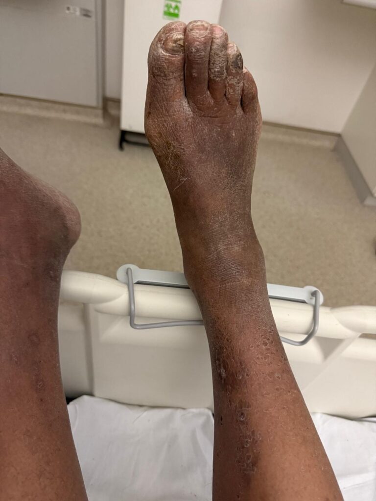
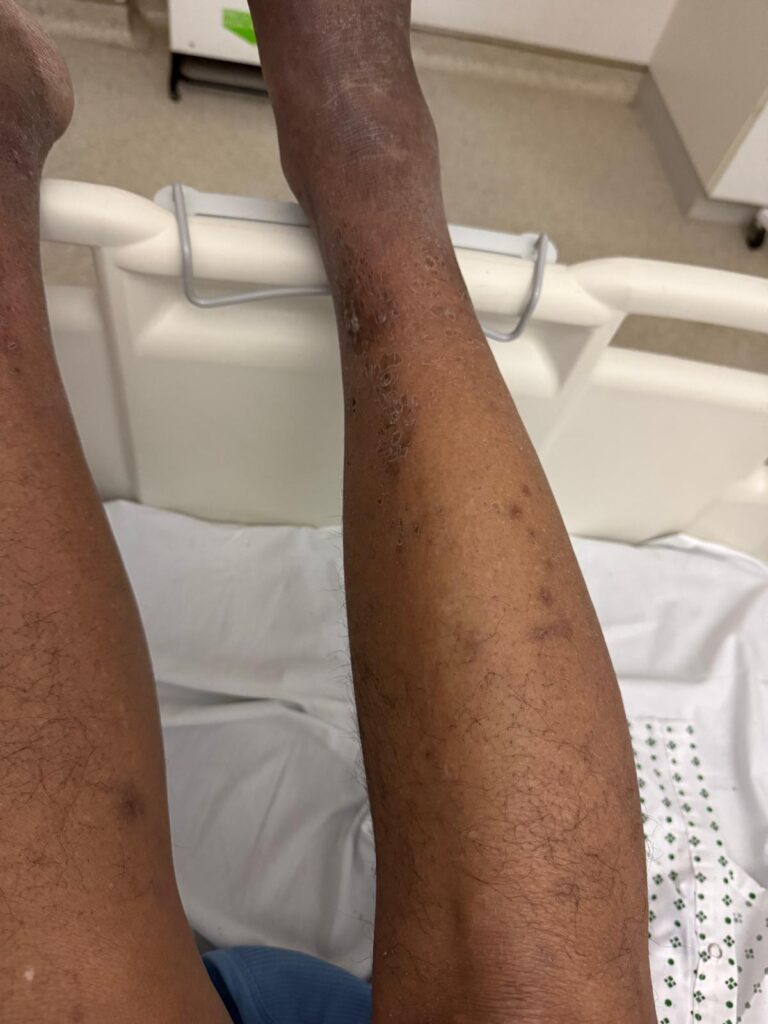
Inspection of the Leg & Ulcer
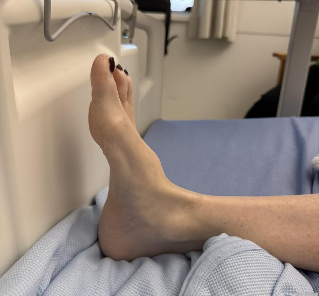
- Count toes — note any amputations; lift foot to inspect heels and lateral aspects; check between toes for infection
- Surrounding skin: lipodermatosclerosis, pallor, dependent rubor, shiny/hairless skin, venous eczema, varicosities
- Ulcer base: colour, depth (is bone visible?), purulent surface
- Ulcer edges: the hallmark of PG — ragged, undermined, violaceous/bluish-red edges; gangrenous (black) margin
- Signs of infection: surrounding cellulitis, erythema, warmth, discharge
- Footwear inspection
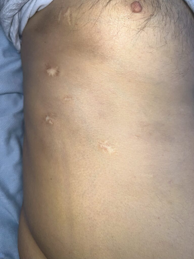
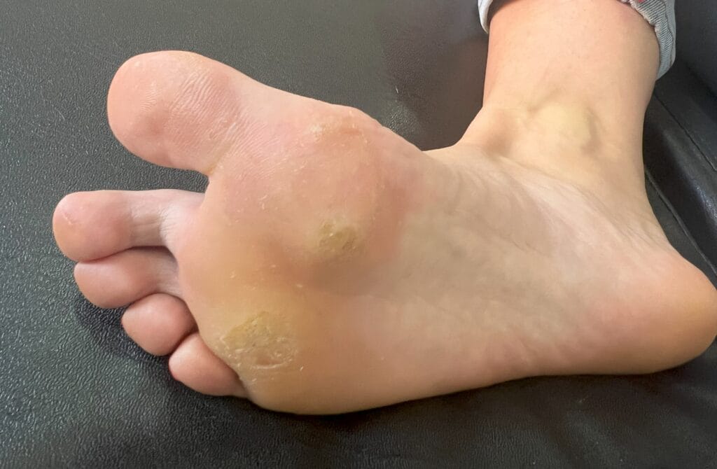
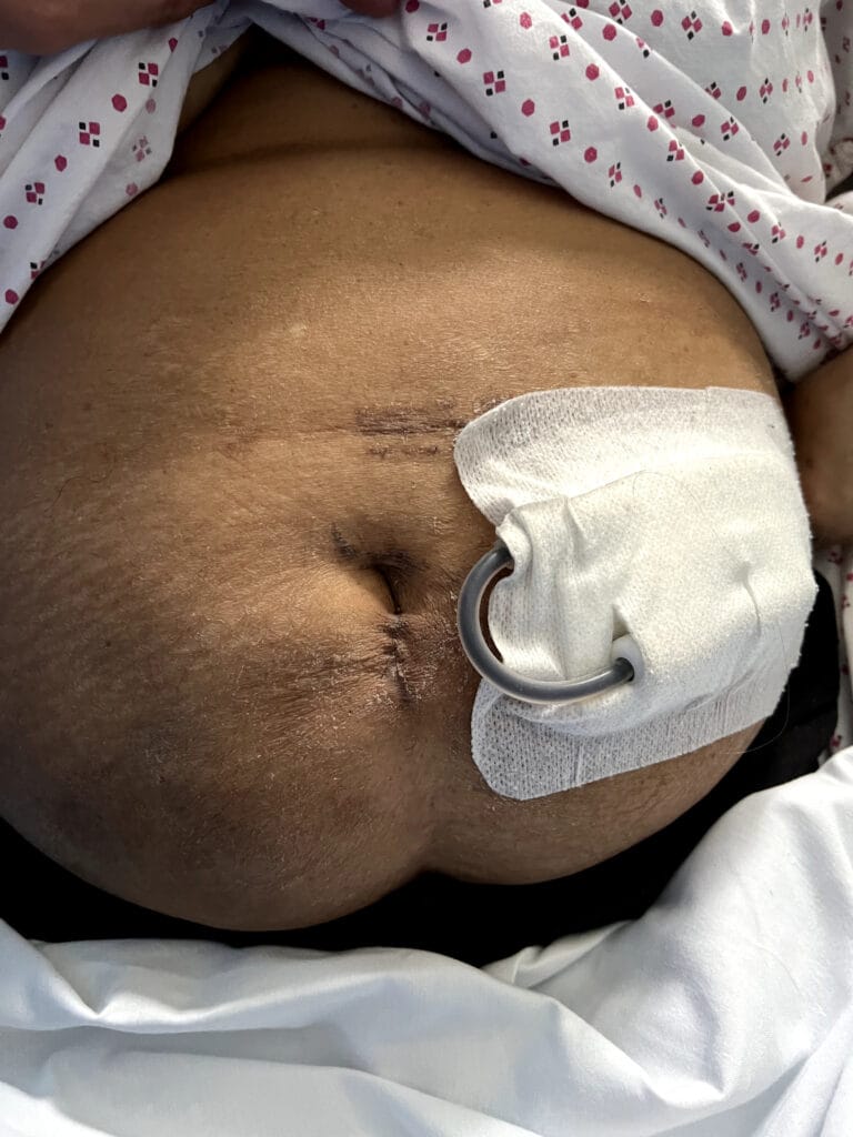
- Stoma sites: do not miss PG around a stoma (a classic PACES trap — PG may koebnerise at surgical sites)
Vascular Assessment
- Temperature gradient and capillary refill
- Peripheral pulses: dorsalis pedis, posterior tibial, popliteal
- Oedema
Neurological Assessment
- Monofilament testing: toes, metatarsal heads, heel, dorsum
- Vibration sense, joint position sense
- Ankle jerk reflexes
Targeted Systemic Examination
- Quick cardiovascular examination (signs of peripheral vascular disease)
- Abdominal examination if IBD suspected
- Lymph node examination if haematological malignancy suspected
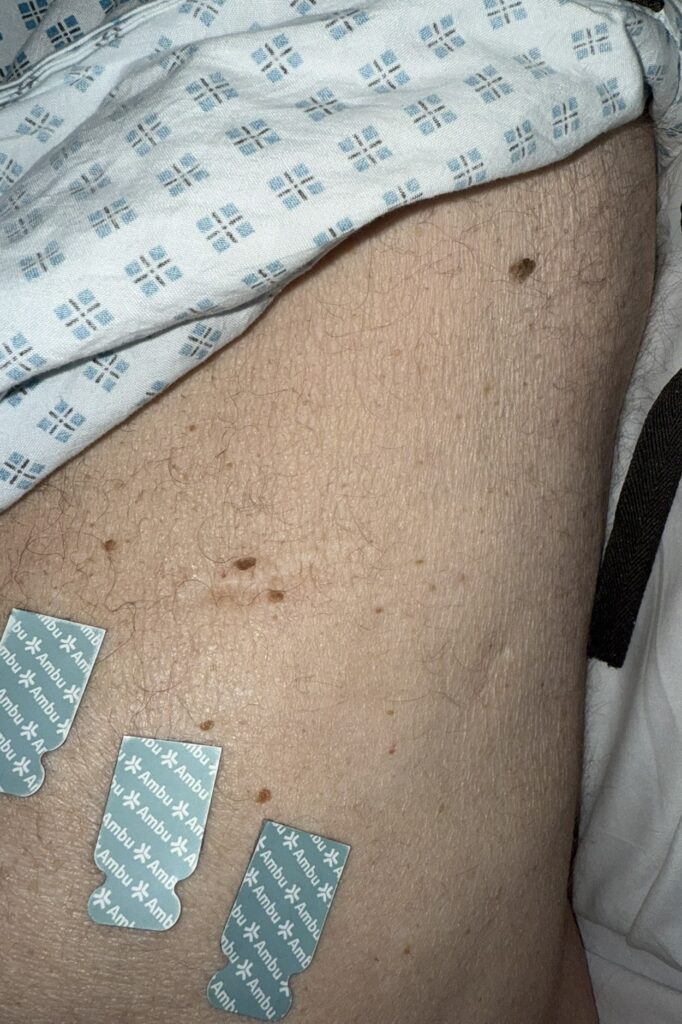
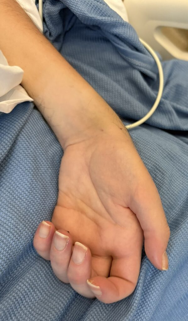
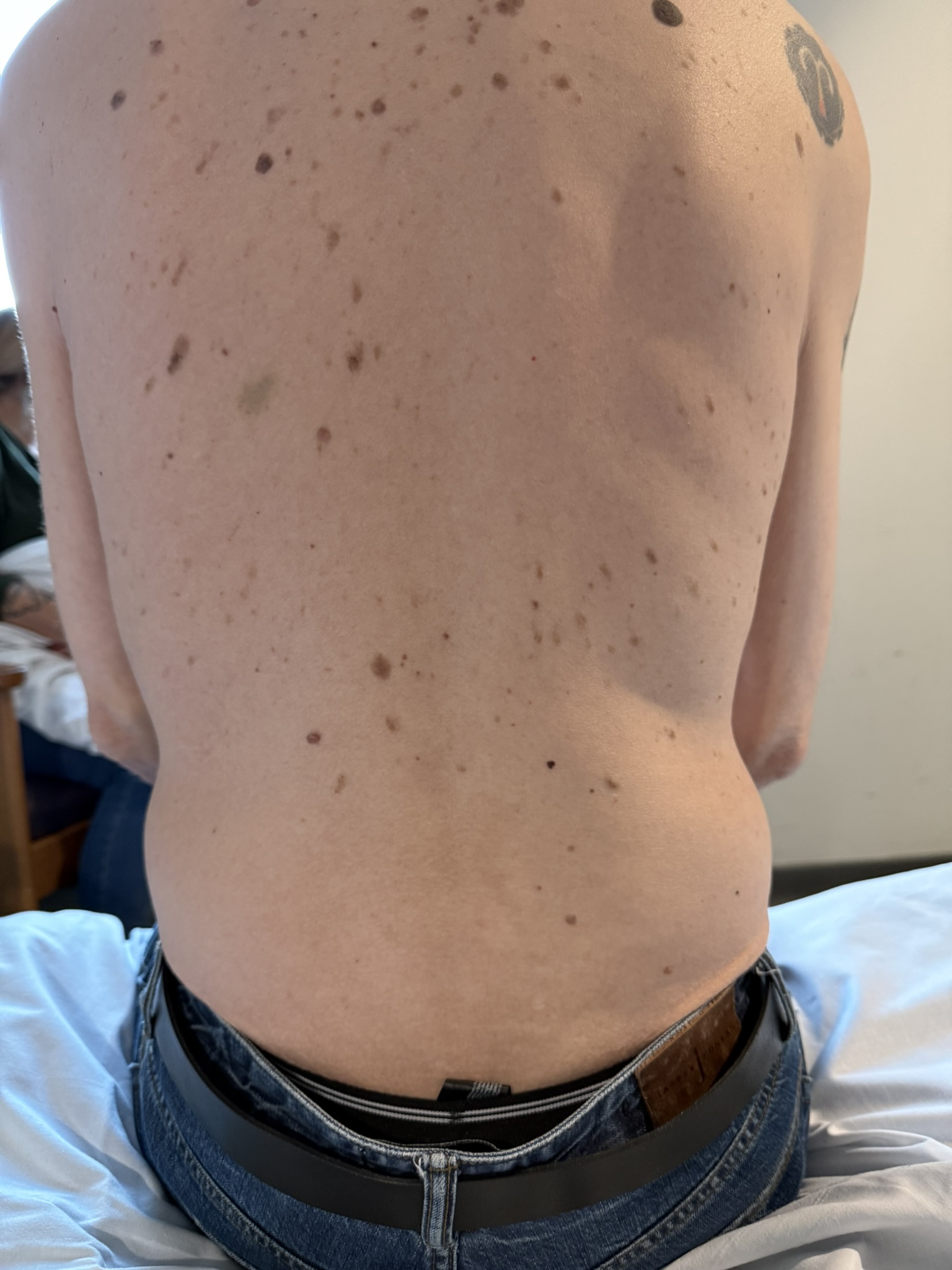
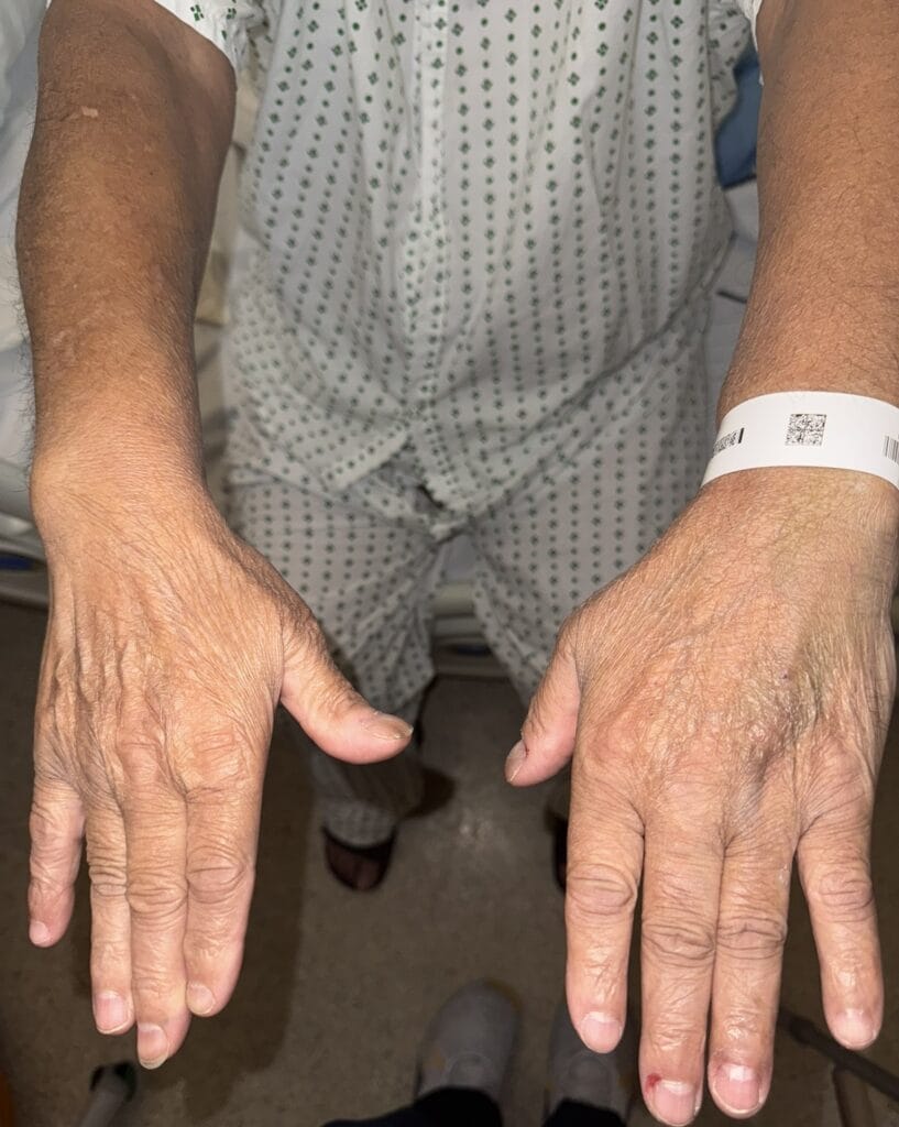
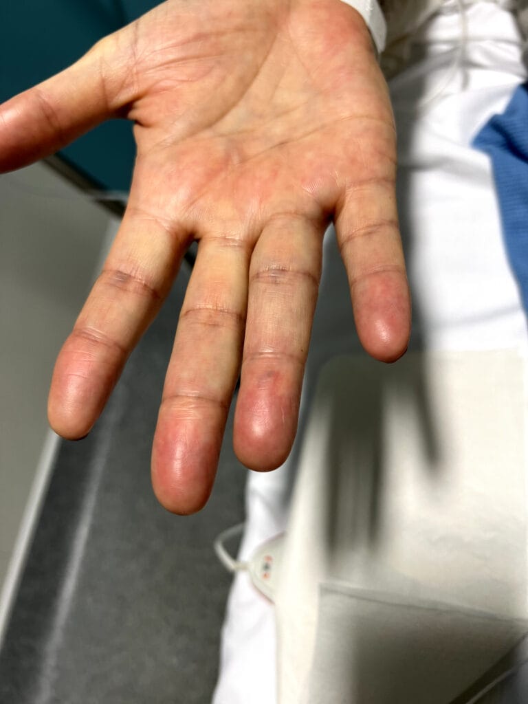
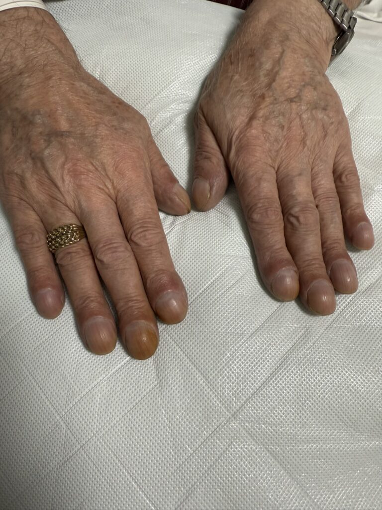
- Inspect rest of skin and hands for arthropathy (RA, seronegative arthritis)
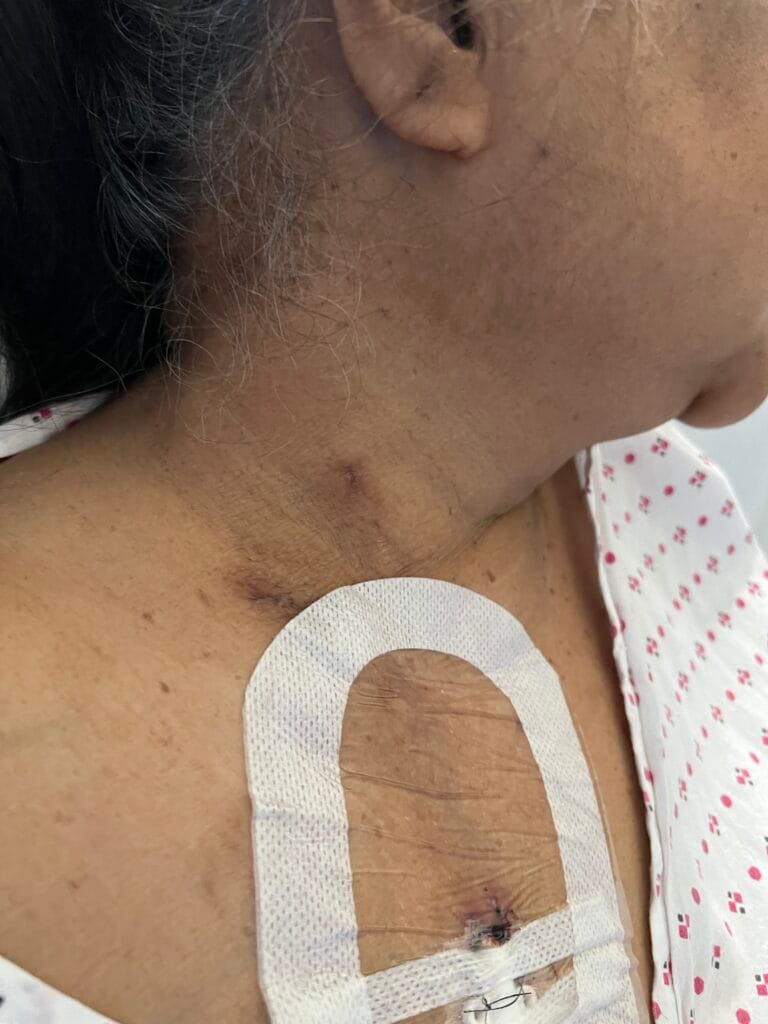
- Medical photography of the ulcer
3. Specific Investigations
Bloods
- Inflammatory markers: FBC, ESR, CRP
- Autoimmune screen: RF, anti-CCP, ANA, ANCA
- Haematological malignancy: immunoglobulins, serum protein electrophoresis, urine Bence Jones protein, calcium
- Hepatic screen: LFTs (to exclude PBC, hepatitis C, autoimmune hepatitis)
- Metabolic: HbA1c, fasting lipid profile (to exclude arterial/diabetic cause)
Microbiology
- Wound swabs — secondary infection is common and must be treated; bacteria isolated do not confirm infection as the primary cause
Vascular
- ABPI (ankle-brachial pressure index) — must be performed before any compression bandaging is applied
Imaging
- X-ray of the limb — to exclude osteomyelitis
- MRI — if osteomyelitis suspected on clinical grounds but X-ray negative
Histopathology
- Skin biopsy — histology is variable and often non-specific (neutrophil infiltrate, epidermal necrosis); biopsy is used primarily to exclude malignancy and other diagnoses rather than to confirm PG. PG remains a diagnosis of exclusion.
Further Investigations Based on Clinical Suspicion
- Colonoscopy if IBD suspected
- Bone marrow biopsy if myeloproliferative disorder suspected
- Medical photography — document ulcer size and appearance at baseline and follow-up
4. Management
- Urgent dermatology referral — immunosuppression and expert wound care are the cornerstones of treatment; do not delay
- Wound care: gentle debridement of necrotic tissue only; avoid wide surgical debridement during the active phase — this can dramatically enlarge the ulcer (pathergy)
- Antibiotics (e.g. flucloxacillin) only if bacteria are cultured or surrounding cellulitis is present — antibiotics do not treat the underlying condition
- Compression bandaging — careful compression can be applied if tolerated and ABPI is adequate, to reduce oedema
- Analgesia — PG is extremely painful; adequate pain relief is essential
- Topical therapies (small ulcers): potent topical corticosteroids, tacrolimus ointment, specialist dressings
- Systemic immunosuppression (larger or refractory ulcers):
- High-dose oral prednisolone (first-line) for several weeks
- IV methylprednisolone for 3–5 days in severe cases
- Ciclosporin
- Anti-TNFα agents (e.g. infliximab — particularly useful if co-existing IBD)
- Mycophenolate mofetil, dapsone, methotrexate, cyclophosphamide, potassium iodide solution
- Treat any underlying associated condition — IBD, haematological malignancy, RA
- ICE discussion: explain the diagnostic uncertainty; outline the need to exclude common causes first (vascular, diabetic, infective); discuss referral to dermatology and the likelihood of immunosuppressive treatment
Pyoderma Gangrenosum Cheat Sheet
| Domain | Summary |
|---|---|
| Genetics / Aetiology | Neutrophilic dermatosis; likely involves dysregulated innate immunity; 50% have an associated systemic disease; remainder idiopathic |
| Epidemiology | Rare; peak incidence age 40–60 years; slight female preponderance; uncommon, chronic, and recurrent |
| Pathophysiology | Dysregulated neutrophil-mediated inflammation; pathergy (koebner phenomenon) is characteristic — new lesions can be triggered by minor trauma or surgery |
| History | Rapidly enlarging, very painful ulcer, often beginning as a pustule or nodule; sudden onset often at site of minor injury; associated IBD symptoms, joint pains, bowel changes, haematological symptoms |
| Examination | Large necrotic ulcer with characteristic ragged, undermined, violaceous/bluish-red edges and purulent surface; most commonly on legs and trunk; check stoma sites |
| Associated conditions | IBD (UC > Crohn’s) ~50%; RA, seronegative arthritis, SLE, GPA, APS; myeloproliferative disorders (AML, CML, HCL, myelodysplasia, MGUS, myeloma); PBC, hepatitis C, autoimmune hepatitis; idiopathic 20–50% |
| Differentials | • Venous ulcer • Arterial/ischaemic ulcer • Diabetic/neuropathic ulcer • Vasculitic ulcer (APS, RA, SLE, GPA, Behçet’s) • Infective ulcer (bacterial, herpetic, syphilitic) • Neoplastic (SCC, cutaneous lymphoma, Kaposi’s sarcoma) • Calciphylaxis, cholesterol emboli |
| Investigations | FBC, ESR, CRP, immunoglobulins, RF, anti-CCP, ANA, ANCA, serum electrophoresis, urine BJP, calcium, LFTs, HbA1c, lipid profile; wound swabs; ABPI; X-ray ± MRI; skin biopsy; colonoscopy if IBD suspected |
| Management | Urgent dermatology referral; avoid wide surgical debridement; analgesia; antibiotics only for secondary infection/cellulitis; topical steroids/tacrolimus for small lesions; systemic immunosuppression (prednisolone, ciclosporin, anti-TNFα, mycophenolate) for larger lesions; treat underlying disease; prognosis unpredictable — may resolve spontaneously or relapse |
← Back to Consultations